Saturday, 28 November 2009
Spot the Dx
Dr Kaur said...
Right Bell's Palsy/ Facial Palsy.
November 29, 2009 10:58 AM
Anonymous said...
Rt Temporal Bone Dysgenesis with Microtia.
November 29, 2009 4:40 PM
dr harpreet s kochar said...
right grade 2 microtia with (most likely congenital) facial paresis and adequate bells phenomenon.
November 29, 2009 10:40 PM
Dr Saumitra Kumar said...
Johnson Mcmillin syndrome.is it?
November 30, 2009 10:44 PM
case of grade ii microtia (Rt)with congenital facial palsy:Dr Nishi Sharma
Looks like cong abnormality of 1st and 2nd arch, need a HRCT temporal bone to llok for other features : Dr Karthikeyan
It is a second arch defect: Dr Sunil Katuria
4th Oman Rhinoplasty and Facial Plastic Surgery course,Muscat, January 12-13,2010
Click here for details
Dear colleagues,
Greetings,
We are pleased to announce the 4th Oman Rhinoplasty and Facial Plastic Surgery course being conducted at Muscat, Oman on 12 and 13th Jan 2010. The seats for hands on cadaver dissection are limited, those interested may please confirm with registration at earliest. The final program with other detail is in attached file. For nonGCC delegates, visa will be arranged.
with best wishes.
Dr.Amar Singh
Course Director
Muscat, Oman.
web: www.entoman.com
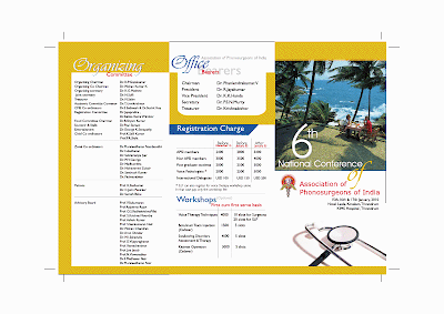
6th National Conference Association of Phonosurgeons of India,January 15-17,2010,Trivandrum
Friday, 27 November 2009
The Lion Live Broadcast-December 9,2009
MENTCON 2009, December 11-13,Nashik
Invitation
Dear Colleague,
Welcome, once again, to Grape City !!!!
We hope you have reserved the dates 11th, 12th & 13th December for MENTCON 2009. On behalf of FMSB of AOI, Nashik Branch of AOI cordially invites you to attend the Annual States ENT Conference to be held at Kavi Kalidas Kalamandir, Nashik.
We assure you, we are leaving no stone unturned in order to make the conference a memorable event................
Conference Secretary
Dr. S. T. Indorewala
Web site http://www.mentcon2009nashik.com
Conference Secretariat
Dr. S. T. Indorewala
Indorewala ENT hospital,
Behind Mahamarg bus stand,
Nashik- 422001.
Ph. +91-253-2580690 Mob. +91-9423173972
Email - mentcon2009nashik@hotmail.com
Sinus Forum Asia 2010 ,Jan 15-17,2010, Kuala Lumpur
Live Diode Laser Surgery Workshops
A series of Lectures and Live Surgeries on Diode Laser are being organised in 8 Cities of India by inviting a Professor from Padua University, Italy. All the details are given in Image Below
Click Image to View
Click Image to View
Sunday, 22 November 2009
Association of Otolaryngologists of India :Joshi Memorial Lecture
Dr Sadashiv G. Joshi ( 1910-1954)
Founder Member of AOI
Graduation 1932(Bombay University) Sandhurst Gold Medal
MS 1934
Chief Surgeon at KEM and Seth G S Medical College, since 1935
Left this World at the early age of 46.His friends and students mobilized donations to start JOSHI MEMORIAL LECTURE in ENT annual Conference of AOI each year.
Most of leading ENT Surgeons from Bombay were his students at that time, who took his work to greater heights later
Click here to read more
Robot-Assisted Endoscopic Thyroid and Parathyroid Surgery with No Neck Incisions.
Thyroid (thyroidectomy, thyroid lobectomy) and parathyroid (parathyroidectomy) surgery has traditionally been performed through a neck incision just above the collar bone. While there have been many refinements to thyroid surgery, patients continue to be unsatisfied with having a cervical (neck) scar.(1,2) By approaching the thyroid or parathyroids through an incision in the axillary fold (under the arm) and using the daVinci Surgical System, we are able to eliminate the cervical (neck) scar and reduce post-surgical numbness of the anterior neck. The recovery time for patients is similar to traditional surgical techniques.
Potential candidates for this new procedure include: patients with thyroid nodules less than 4 cm in diameter, some papillary thyroid carcinomas, and some follicular thyroid carcinomas. Additionally, patients with a parathyroid adenoma that can be localized pre-operatively through imaging studies may be candidates.
Click here to see video
VISIT http://www.roboticthyroid.com/home
Saturday, 21 November 2009
Combined approach technique for the management of large salivary stones.-abstract
Walvekar RR, Bomeli SR, Carrau RL, Schaitkin B.
Department of Otolaryngology and Head and Neck Surgery, University of Pittsburgh Medical Center, Pittsburgh, Pennsylvania 15213 , USA.
OBJECTIVES/HYPOTHESIS: To review our experience with the combined approach, which includes an internal (endoscopic) and open approach for the management of large salivary stones. STUDY DESIGN: Retrospective. METHODS: Clinical data was reviewed on patients who underwent combined approach for large salivary stones from July 2005 to August 2008 under an institutional review board approved protocol. RESULTS: Of 106 patients who underwent sialendoscopy, 19 patients (18%) had 20 combined procedures. Thirteen patients (68%) were women and six men (32%) with a mean age of 52 years (range, 15-69 years). Operative times were shorter for submandibular stones (mean, 90 minutes) as compared to parotid stones (mean, 133 minutes). Stone removal was successful in 90% (18/20) with no major complications and minor complications in four patients (20%). CONCLUSIONS: The combined procedure allows complete removal of large or impacted sialoliths without the need for removal of the entire gland with acceptable complication rates. We also recommend consenting patients for this approach when a difficult endoscopic removal is anticipated. Laryngoscope, 2009.
PMID: 19358166 [PubMed - indexed for MEDLINE]
(One such live surgery for Parotid duct stone was done at a course at GTB Hospital,Delhi by Dr Francis Marchal from Geneva on 21.11.2009)
Otolaryngology Poll
What single factor dominates Otolaryngology today?
1.Skill
2.Knowledge
3. Technology
VOTE AT www.entindia.net
1.Skill
2.Knowledge
3. Technology
VOTE AT www.entindia.net
Friday, 20 November 2009
CIGICON 2009, Photographs
Here we are providing the links to view conference photos
Pre conference and Faculty Dinner photos: plz copy and paste the following link in address bar
http://picasaweb.google.co.in/cigicon2009hyd/CIGICON2009PreConference?feat=directlink
Main Conference Photos:plz copy and paste the following links in address bar
http://picasaweb.google.co.in/cigicon2009hyd/20091118?feat=directlink
http://picasaweb.google.co.in/cigicon2009hyd/20091119?feat=directlink
--
CONFERENCE SCRETARIAT
7th Annual Conference CIGICON-2009
ENT Department, Apollo Hospitals, Apollo Health City Campus
Jubilee Hills, Hyderabad-500033
Ph:+91-40 2360 7777 Ext.3737
Fax: +91-40-2360 8050
Email: cigicon2009hyd@gmail.com
Website: www.cigicon.org
Saturday, 14 November 2009
International Course on "Sialendoscopy" November 20-21,2009
International Course on "Sialendoscopy"
Venue: UCMS & GTB Hospital ,Delhi
Dates : November 20-21, 2009
Course Director: Dr P P Singh
Professor & Head, Deptt of ENT,
UCMS & GTB Hospital,
Dilshad Garden, DELHI - 110 095
Guest Faculty : Dr Francis Marchal
Director (ESTC),
European Sialendoscopy Training Centre,
Geneva (Switzerland)
Course Faculty : Dr Arun Goyal
Reader, Deptt of ENT,
UCMS & GTB Hospital,
Dilshad Garden, DELHI - 110 095
Contact: arungoyal150@hotmail.com
Mobile: +91 11 9810812813
Website: www.otologyindia.blogspot.com
Frontal 2009, Kochi 2009, November 20-21,2009
Frontal 2009
( International workshop on Frontal Sinus surgery, 20th & 21st November 2009)
Welcome back to Kochi!
We are here again with an exclusive in depth demonstration of the most difficult region in the sinuses, THE FRONTAL, led by none other than the doyen of the frontal sinus, Prof.Wolfgang Draf, the person who described the Draf type I ,II,& III frontal sinus drainages.
Prof.Draf, and Dr.George Varghese will spend two days demonstrating live frontal Sinus surgeries. Well, one cannot finish the FESS by just entering the frontal Sinus alone! It goes without saying that in addition to the live demonstration of the different frontal Sinus drainages, routine FESS will also be shown.
So, gear up, join for this first of its kind course. GV ENT Centre, & ‘The Advanced Rhinology Centre’ at the Vijayalakshmi Medical Centre will not leave any stones unturned, to make every minute you spent with us worthwhile. Please note that we can accommodate only 300 delegates, & so register now, so as not to get disappointed.
click here to download Registration Form
Click here to download brochure
See you in November 2009.
Your’s Sincerely,
Dr.George Varghese Dr.Saju KG
Course Director Org.Secretary
CONTACT 09447407706
EMAIL
georgegogi@gmail.com
www.gvent.net
( International workshop on Frontal Sinus surgery, 20th & 21st November 2009)
Welcome back to Kochi!
We are here again with an exclusive in depth demonstration of the most difficult region in the sinuses, THE FRONTAL, led by none other than the doyen of the frontal sinus, Prof.Wolfgang Draf, the person who described the Draf type I ,II,& III frontal sinus drainages.
Prof.Draf, and Dr.George Varghese will spend two days demonstrating live frontal Sinus surgeries. Well, one cannot finish the FESS by just entering the frontal Sinus alone! It goes without saying that in addition to the live demonstration of the different frontal Sinus drainages, routine FESS will also be shown.
So, gear up, join for this first of its kind course. GV ENT Centre, & ‘The Advanced Rhinology Centre’ at the Vijayalakshmi Medical Centre will not leave any stones unturned, to make every minute you spent with us worthwhile. Please note that we can accommodate only 300 delegates, & so register now, so as not to get disappointed.
click here to download Registration Form
Click here to download brochure
See you in November 2009.
Your’s Sincerely,
Dr.George Varghese Dr.Saju KG
Course Director Org.Secretary
CONTACT 09447407706
georgegogi@gmail.com
www.gvent.net
Otolalaryngology Pool: What do you prefer to call your self as ENT Professional?
1 ENT Specialist
2 ENT Surgeon
3 ENT Consultant
4 Otorhinolaryngologists
5 Otolaryngologists-head and neck surgeon
Vote at www.entindia.net
Comment welcome
2 ENT Surgeon
3 ENT Consultant
4 Otorhinolaryngologists
5 Otolaryngologists-head and neck surgeon
Vote at www.entindia.net
Comment welcome
Turkish delight
Versatility of diced cartilage-fascia grafts in dorsal nasal augmentation
Authors: Martin H Kelly, Neil W Bulstrode, Norman Waterhouse
Journal: Plastic and reconstructive surgery
BACKGROUND: Augmentation of the nasal dorsum using autologous cartilage remains an exacting task in rhinoplasty. Precise, long-term control over graft contour and alignment can be difficult to achieve. In an attempt to alleviate these problems, the use of diced cartilage wrapped in a supportive sleeve has recently seen a resurgence of interest. The Turkish delight technique uses Surgicel as the sleeve material, whereas Daniel and Calvert have proposed autologous fascia. METHODS: The objective of this study was to assess the efficacy and reliability of diced cartilage-fascia grafts in the treatment of dorsal nasal volume deficiencies. RESULTS: Over a 2-year period, 20 adult patients underwent nasal augmentation using diced cartilage-fascia grafts. The indications for dorsal grafting were congenital saddling, racial refinement, or iatrogenic or posttraumatic deformities. Apart from one infection, all of the hybrid grafts retained their original volume and had not undergone resorption by a mean time of 16 months postoperatively. A critical analysis of the radix-dorsum aesthetics led to a minor modification of the original technique. CONCLUSION: This series supports the use of diced cartilage-fascia grafts for the correction of difficult dorsal nasal defects with grafting in the 3- to 5-mm range in primary and secondary rhinoplasty.
Plastic and reconstructive surgery. 01/12/2007; 120(6):1654-9; discussion 1654-9.
ISSN: 1529-4242
DOI: 10.1097/01.prs.0000285185.77491.ab
PS A modified technique using fascia lata instead of surgicel was demonstrated by Dr Virender Ghaisia, Pune today in country -wide teleconferencing sponsored by CIPLA Respiratory
Friday, 13 November 2009
CT evaluation of anatomical variations of osteomeatal complex:Indian Journal of Otolaryngology &HNS
The main article in July-September 2009 issue Indian Journal of Otolaryngology &HNS
Authors :AA Wani Sohit Kanotra M Latif et al
GMC Srinagar
A total of 150 patients of chronic rhino sinusitis (medical treatment failure ) who were subjected to FESS were CT scanned pre operatively to find any anatomic variation and extent of mucosal disease
Concha Bullosa was commonest anatomical variation and was seen in 45 (30%)patients. The others variations included :paradoxical middle turbinate in 9.33 %patients ,uncinate process variation in 25 % patients, agar nasi cell in 9.33 %,Haller cell in 8.66 % and posterior septal deviation in 25.33 % patients
The mucosal disease was most commonly seen in anterior ethmoids (87.33%),followed by maxillary sinus ostial area (70 %),posterior ethmoid disease (38%),frontal sinus disease (15 %)and sphenoid mucosal disease (8.66%) patients .
The authors have tabulated the prevalence of anatomical variation of other few studies ,each showing different percentages.
Another analysis has been done on involvement on one side or both sides. Where as most anatomic variations are predominantly unilateral but most mucosal disease is bilateral (except frontal and sphenoidal
The authors conclude that pre operative detection of various anatomical variations is essential as it significantly influence the selection of technique and also helps in avoiding complications .
Wednesday, 11 November 2009
Pemberton's sign.
Movie clip at youtube
http://www.youtube.com/watch?v=laa81LS2Oho
Dr Harpreet S Kochar
Consultant
Dept of ENT and Head Neck Surgery
Kailash Hospital
Greater Noida
India
Web: www.entgreaternoida.com
http://www.youtube.com/watch?v=laa81LS2Oho
Dr Harpreet S Kochar
Consultant
Dept of ENT and Head Neck Surgery
Kailash Hospital
Greater Noida
India
Web: www.entgreaternoida.com
Tuesday, 10 November 2009
Belly Dance of an Ant - Dr Janki Ram ,Trichy
click here to see video
Please keep sound on
More ENT Videos FROM DR JANKI RAM
WWW.YOUTUBE.COM
ID : DrJanakiram123
ENT ENCYCLOPEDIA
Novel totally implantable piezoelectric device
"A novel totally implantable piezoelectric device has been developed by Envoy Medical Corporation, USA. This device, Esteem-Hearing Implant is 100% implantable, so it is 100% invisible. Esteem has a piezoelectric transducer (Sensor) placed on the Incus bone which converts the mechanical energy into electric energy, which is the known property of a piezoelectric material. This electrical signal is then amplified, processed and converted back to vibratory signal by means of another piezoelectric transducer (Driver) placed on stapes. The piezoelectric transducer has been shown to provide output levels close to 110 dB SPL. Like a hearing aid, an Audiologist programs the implant using a device called commander and once programmed, patients have the option of adjusting volume, modify background noise filters and turn it off/on remotely".
Please Visit www.AudiologyIndia.com for more information and clinical report.
Saturday, 7 November 2009
Hands on Workshops in India -Contribute information
Can you please give some informations of :
1) temporal bone dissection workshops held and conducted in India ?
2) FESS cadaveric dissection courses conducted in India ?
Regards
Dr.JAYANTA KUMAR NATH MBBS,MS(ENT)
C-41,Jal Vayu Vihar,
Sector III,LB Block,Salt Lake,
Kolkata 700098
West Bengal, India
Send information to update@entindia.net for compilation
or you can put information in comments below
Supracricoid laryngectomy
Supracricoid laryngectomy includes removal of the entire supraglottis, the false and true vocal cords, and the thyroid cartilage including the paraglottic and preepiglottic spaces. In addition, one arytenoid may be resected. The cricoid cartilage, hyoid bone, and at least one arytenoid are saved. Phonatory and swallowing function is maintained by the movement of the spared arytenoid against the tongue base. Respiratory function is dependent on the preservation of the cricoid cartilage. Preservation of these laryngeal functions form the basis of conservation(organ-preserving) surgery.Click here to read more
Botox Treatment for Vasomotor Rhinitis (Idiopathic or Non-Allergic Rhinitis)
Nasal hypersecretion due to idiopathic rhinitis can often not be treated sufficiently by conventional medication. Botulinum toxin A (BTA) has been injected into the nasal mucosa in patients with nasal hypersecretion with a reduction of rhinorrhea lasting for about 4 to 8 weeks. Since the nasal mucosa is well supplied with glands and vessels, the aim of this study was to find out if the distribution of BTA in the nasal mucosa and a reduction of nasal hypersecretion can also be reached by a minimally invasive application by sponges without an injection.More....
Wednesday, 4 November 2009
www.audiologyindia.com
Greetings from www.AudiologyIndia.com
We are aware of your Blog on ENT related topics and it is one of the best regularly updated blog and with lots of infomation.
Many thanks
Best regards
AudiologyIndia.com Team
Otolaryngology Poll-Voice your Opinion
Are the Academics in National Conference shadowed by tourism, cocktails, exhibitions & politics?
1 Yes
2 No
3 Couldn't care less !
Vote at www.entindia.net
1 Yes
2 No
3 Couldn't care less !
Vote at www.entindia.net
International Pediatric Otolaryngology conference,Hyderabad ,March 13-14,2010
We at Hyderabad are coming out with International Pediatric Otolaryngology conference from March 13-14, 2010, under the banner of AOI-Hyderabad in association with Association of Pediatric otolaryngologists of India. In this regard I am attaching a conference flyer for display in the ENT update website. I shall be grateful for the favor and shall be glad to answer any queries. Look forward to seeing you in the upcoming conference at Hyderabad in March 2010.
Best regards,
Sincerely
Vishnu Vardhan Reddy Martha
M.S.,DLO,DNB,Ph.D
PRESIDENT AOI-HYDERABAD
ORGANISING CHAIRMAN
INTERNATIONAL PEDIATRIC OTOLARYNGOLOGY
CONFERENCE, MARCH 13-14,2010
VENUE: HYDERABAD.
Professor of Otolaryngology,
Osmania Medical College,
Hyderabad-90,
Andhra Pradesh, India.
E-mail: vishnumreddy@yahoo.co.in
Tuesday, 3 November 2009
Use of GV Paint in ENT
Dr Ajit Man Singh writes
GENTIAN VIOLET PAINT was reccomended to me by my teacher on one post op myryngoplasty, where the repaired drum refused to dry up, and remained congested, despite anatomical closure.
I have since used it extensively in the ear: post tympanoplasty, mastoid cavities, otitis externa, otomycosis also, one teache erccomended for apthous ulcers, where also I have used.
I find that it does wonders.
however, I do not have any evidence of adverse effects, except fot the drycleaning bills of ones shirt.
Would appreciate inputs from others.
Dr N N Mathur Writes:
It is in fact an old and trusted paint. I never used it in ear post myringoplasty/mastoidectomy, but I have extensively used it on some small fistulas/defects that remain in neck following head neck surgery. It does work vey well. Fortunately such defects are less now with better radiation/ cautery/ antibiotics and postop care. Only problem with it is difficult assessment of the site post application.
Dr Anil Safaya (Oman)Writes;
We used to use GV paint extensively in cases like apthous stomatitis and I agree, the results used to be astounding !!!...I agree, I am also keen to know more inputs!!!
Blog Author Comments:
One study has linked long term exposure to large amounts of Gentian violet with cancer. The Food and Drug Administration in the US has determined that gentian violet has not been shown by adequate scientific data to be safe for use in animal feed. Use of gentian violet in animal feed causes the feed to be adulterated and is a violation of the Federal Food, Drug, and Cosmetic Act in the US. On June 28, 2007, the US food and Drug Administration issued an "import alert" on farm raised seafood from China because unapproved antimicrobials, including gentian violet, had been consistently found in the products.Read more
Monday, 2 November 2009
Spot the Diagnosis
The cytology shows multinucleated giant cells
Please use Comments logo below
venkat said...
dentigerou cyst
November 1, 2009 11:48 AM
Anonymous said...
?hemangiopericytoma
November 1, 2009 7:07 PM
Anonymous said...
ca maxilla....bulge in maxillary sinus area besides the bulge in the hard palate
November 1, 2009 7:51 PM
Dr. Vijay Sharma said...
The nodular lesion may be because of leprosy.
Dr. Vijay K Sharma
November 1, 2009 9:29 PM
Dr. Dheeraj Mishra said...
Maxillary Fibro-Osteoma
November 2, 2009 12:38 PM
saumitra kuamr said...
tuberculous ostemyelitis of the hard palate(maxilla)
November 2, 2009 7:16 PM
DR ATUL KANSARA said...
ectopic salivary gland tumour[may be pleomorphic adenoma]
DR ATUL KANSARA
AHMEDABAD
November 2, 2009 10:40 PM
anil said...
Multineucleated Giant cell is the typical feature of Tuberculosis.
This is a case of Tuberculosis of alveolus of tooth.
Dr Anil Jain
Delhi
November 3, 2009 9:14 AM
Anonymous said...
ectopic salivary gland tumour[may be pleomorphic adenoma]
November 3, 2009 2:53 PM
Subscribe to:
Posts (Atom)